
Day 5 - Human Dissection in Phoenix
After some R&R from this highly intense week, here's the fifth installment of my human dissection experience! I think of all the days, this might be the most interesting. There was a lot of things going on, people working on the last parts and so many questions. And brains!
We started the day off with completing the evisceration. So we first had to detach the pectoralis from the chest, and take off as much of the diaphragm as possible from the "roof" of the chest cavity. After that we could start cutting the ribs on both sides. We took turns cutting the ribs - they do have a certain feel to them. We used common gardening cutters, the same you would prune your fruit trees with. Certainly an unusual feeling to use them for ribs. The ribs were fragile and porous, they had a pinkish tone and looked like there were bubbles in them. You could almost hear the texture as you cut the rib, it wasn't a "hard" crack, but rather like breaking it apart type crackle. To finish removing the chest plate, we cut right under the clavicles across the chest. The chest plate was beautiful from the inside, lots of muscle in between each rib, different tissue directions that could be seen under the shiny membrane covering them. Very cool! At last we could see the lungs and the sack for the heart! I was quite surprised to see that the heart was placed so low in the body, because from the pictures you see in a typical anatomy book, it's placed quite high. There are probably individual differences (as with all else), but still.
Good to know that even though anatomy books provide us with a map, the terrain isn't always the same.
From there it was quite easy to detach the diaphragm from the back and spine. It really does have a lot of attachments! And what fascinated me most about the diaphragm was that it was SO thin. It almost felt plastic bag type thin (though it probably was a bit thicker at least). I'm surprised by plenty of tissue in the body, how can something so thin be so strong and durable? How can we abuse things so, and still they do their job just fine? I think it's thinness means that it does a good job moving though. When you think about it a strong and thick diaphragm would probably mean less ease of breathing, and because it's attached to so many other things, most likely that it would inhibit movement across the whole body.
After detaching the diaphragm, it was time to remove the heart sack, with as much surrounding vessels as possible. It wasn't easy! Both the sack and the heart itself had plenty of yellow fat on it. You wouldn't think so, but I guess it's pretty good for keeping the heart safe. I'm not sure if it was a normal or unhealthy amount of fat, I forgot to ask. But when looking at our cadaver on day 1, there was no reason what so ever to believe she was overweight. And her greater omentum was so small, that I think she might not have had enough fat in the right places. But I don't know!
It was a surreal feeling to take out the heart and hold it in my hands. The aorta alone, is as big as the circle I can make by putting my thumb and index finger together! And it is long too! I'm struggling to understand the volume of blood that goes through the heart in only a minute. And how strong and wonderful the heart is to keep doing it's job. Dissecting the heart was really difficult, we weren't sure were to start, or finish. It was a big mess of muscle, fat and vessels. Nonetheless, amazing!
After finishing up the heart it was time to get the lungs out. I wanted to make a good model for us to study outside the body, so I removed the trachea together with both lungs. I also managed to get the thyroid membrane and cartilage, and anything in between that and the trachea. It was hard work getting it all out in one piece, but totally worth it once completed. They actually looked nicer outside the body than on the inside. Our cadaver had some strange lumps on the front of her lungs, which turned out to be some sort of repair of non-efficient alveoli (I think). I can't remember the name of it, but if I recall correctly it was that if two alveoli doesn't function well, they can grow together to be able to regain the function that was previously lost. I'm gonna have to do some research so I don't forget about that completely! Did you know that the alveoli makes up for about 70-75m2 of surface? Crazy, right?
The lungs had an extraordinary texture, it was like minimi bubble wrap to touch. Almost as if you could feel the air going out them! It reminded me of the texture of the aponeurosis on the skull. Very strange, yet fascinating. The color was a deep red/brown. Were the trachea went into the lungs, it felt like bigger bubbles, almost like what the artery plaque felt like, but I'm guessing it's supposed to feel that way there, considering the thickness and feel of the trachea. It was pretty much a tube! Going almost all the way around, but it was soft to the touch against the back. The tube feel went all the way from the insertion in the lungs, to the thyroid membrane.
We took the chance to look closer at the discs when we had freed up the chest cavity. The discs have a really interesting texture and feel to them. They were not at all as soft as I would have imagined. And they looks like trees when you cut them up, with rings going around it. Considering the tightness of everything around the spine, it's amazing that we can even get herniated discs. I wonder how many painful cases of herniated discs and bulging discs that are actually caused by the tissue around the spine and what's supporting it (like the hip), rather than the discs. And I can see how movement would still be beneficial when herniated, as the tissue around the herniation would likely be able to help everything come back to its right place.
Anyway.
When done with the chest cavity, it was time to move on to a different cavity, at the opposite end of the body. I hadn't finished uncover the pelvic floor on Thursday, so this was the time to finish the job. It was a pain the the butt (we even had some slight leakage from said butt). It was almost impossible to do a good job, so I got the bladder, uterus, sigmoid colon/rectum and the whole outside to go in one piece.
What happened next was probably my least favorite part of the week - it was so out of this world and just plain weird! Todd came by and asked about the uterus, which was intact and could be displayed. We also found the Fallopian tubes and ovaries. Todd then asked me to find the cervix, which wasn't all that hard. And to see it, I was to push it out through the vagina. This is were most of the butt leakage happened, along with great difficulty getting the thing out! We had to cut the opening to be able to see anything (I certainly feel my fiffi clam up by then!), and when not even that was a success we (finally) got to cut it up from the inside. This was just the weirdest thing ever! But at least now I know how to tell if someone has had babies or not. If someone has not had a baby, the opening of the cervix is round, and if someone has had baby, the opening changes for to an oval opening, or a line. So I'm glad to say our cadaver had the chance to reproduce while she was alive! The cervix was actually out to the test by two other students, I think they could only open it a few millimeters, which makes it really difficult to understand THATS WERE BABIES EXIT !
After that vaginal adventure, I had to focus on something that wasn't in the lower part of the body. So, me and Johan quickly removed the temporalis on both sides, to prepare for a proper, real life brain scan.
Todd came up with an electric bone saw, me and Oscar held the head and Todd did the hard work. He went all the way around the head, almost all the way through. Cranium smells just like when drilling in teeth by the way. Before going any further, he asked one of the students to grab one of the red tissue bags and turn it inside out. This was for catching the brain! He then proceeded to put a flat screwdriver in between the sides of the cranium and hammer it off. That sound, of bones tearing, was a bit creepy... And like a helmet, the cranium was off! Me and Oscar held the cadaver up the whole time, to keep the brain from spilling over. It really had NO shape what so ever! You couldn't tell one part from the other(it did have the characteristic folds though). So we then went on to pour the brain onto the red bag, what a mess... Inside was still the cerebellum, some of the cranial nerves attached to it and the nerves going to the eyes. We poured that too. I had brains up to my elbows after this, I never thought I'd say that 😳
An embalmed brain would have a quite different texture, more like rubber. That would really help if one would like to study the brain up close, as now we missed "all the fun", as the brain just melted out without any specific part being firm enough to make out.
One of the other groups opened their cranium a bit differently, the first took of the mandible and then used a handsaw to go up the head at an angle. That created three lovely pockets to study after pouring out the contents. A fantastic view!
After seeing the brain floating out like that (twice!) I can't believe how it can actually function! And I'm amazed by the fact the more people doesn't seriously injure their brains while doing stupid and/or crazy things! Also intrigued by the causes of epilepsy (which I have), how big or small is the impact? How much damage does my brain suffer for each of my seizures? I hope I don't have to have any more of those.
One thing that was interesting about the two skulls was the difference in thickness of the cranium. It ranged from about 3-6 mm in one of them (male) to 6-10 mm in the other (female). Ours, the female, also had necrosis of the bone by the frontal lobe. That makes you think about whether or not she had any behavioral problems before she died. Quite interesting really. Apparently the cause of this type of necrosis isn't yet known.
After finishing up with the brain I went to see what Oscar was up to with Todd at one of the other tables. They were dissecting an eye and found an artificial lens! So turns out that cadaver had previously had eye surgery, perhaps for glaucoma or something similar. We also got to see a jelly like piece of tissue that "lives" in the eye. Can't remember the name, but it was fascinating.
After all the head action I went to go through the foot with Staffan. What a beautiful creation. No wonder the foot can cause problems, it's really tight and well planned down there to start with, so any loss of movement or an injury would definitely have consequences! Think of when you go to IKEA and see one of their tiny bedrooms, all well planned and perfect. But if that was your home, there would probably be laundry, books and other things lying around the whole place making the space a lot less functional for you. That is your feet.
Other findings during the day was that our cadaver had a bone spur both in the subtalar joint, which made dorsal flexion of the foot impossible. There was also a bone spur in the acetabulum in one of the hips. A bone spur is pretty much bone tissue where it shouldn't be, usually right in your joint. Don't grow them, they are likely to hurt! Our cadaver also had scoliosis, probably acquired through lifestyle in Todd's opinion. I wouldn't know how to tell the difference!
Another thing we reacted to was how small the thoracic vertebrae is compared to the lumbar vertebrae. I would say even more than in the typical models we use in school.
After lunch we had a look around all the other tables to learn more about everybody else's findings. I learned how small and tiny the meniscus is, and that the bursa pretty much looks like fat. I would have missed it if it wasn't pointed out to me! We also took a good look at the ACL, which two of the guys tried to tear. In just flexion it was pretty much impossible, but with some knee valgus it came off a lot easier! Again, another horrible sound that I'll probably remember forever.
One of the other tables had a woman who had been to surgery and got two big tubes inserted in her body, going from the heart, all the way down to the femoral arteries of both legs. All in all it was about 2.5m of plastic tubing, encased by her body. Extraordinary. As they removed it, they found it went well down into her thighs. We're not sure of the reason why this procedure was done. Do you have any idea?
The woman with four toes (eight all in all) actually had two screws in each of her foot. There wasn't enough time to find out more, but the group working at the table reasoned that they might be there to give the feet a bit more stability, which might be lacking with only four toes. Unfortunately we will never know, but it's interesting to speculate at times.
We also had the opportunity to closely inspect the penis and testicles. I can't remember everything that was said about it, but one of the things that came up was erectile dysfunction and what may cause it. One reason is bad blood circulation, which may make it harder for the blood to make its way into the spongiosus-thingies. There's also the pudendal nerve that can be impinged when sitting a lot. It lies right in between the sacrotuberous ligament and the sacrospinous ligament, so it would quite easily get impinged if those two where too tight.
Other interesting facts you may be interested to know is that the way you expand skulls for education (where the parts of the cranium comes apart), is to insert a tight bag of rice into the head, and to then book it. Viola, cracked skull! Also, the best way to remove tissue from bone is an insect bath (apparently all big institutions has one), that will clear bones of any edible tissue. If the bones need to be whiter they can then be bleached.
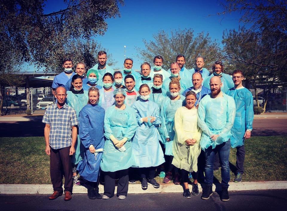
After the day we had a short break to thank Todd for the week, and give him a gift as a token of our appreciation!
Then it was time to clean up and prepare the bodies for their journey home to their families. They were all put together with their body part and tissue in a plastic bag to be cooled down. We then got to help out with taking out ten frozen cadavers that had already been dissected for thawing over the weekend, as they too were to make the journey home for cremation and burial with their families.
Thank you for reading! :)